MRI Requirements: ASHS-PMC-T1 Atlas
Overview
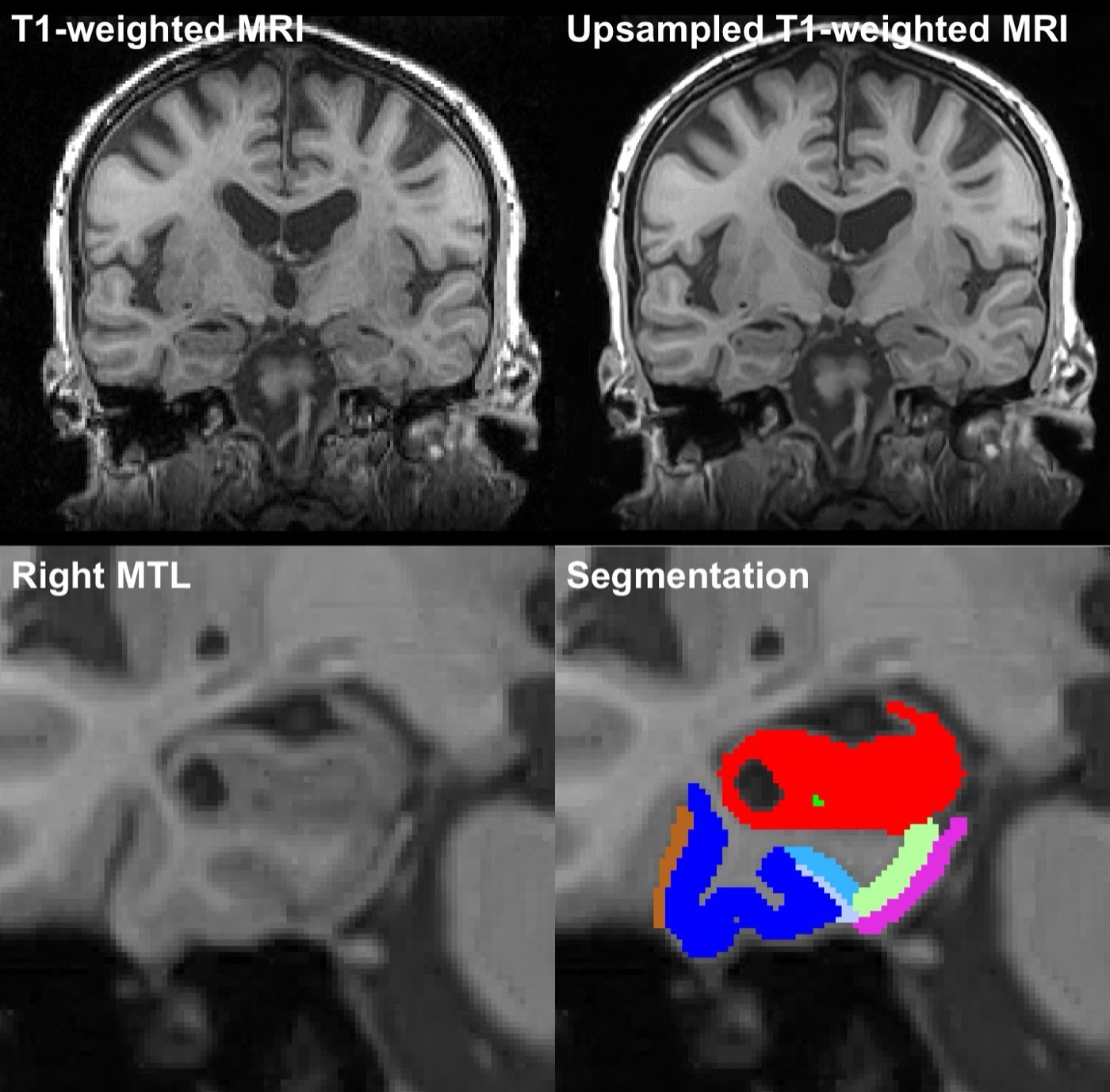
This page describes the MRI data requirements for using ASHS with the ASHS-PMC-T1 atlas. This atlas is used to automatically segment the whole hippocampus (anterior and posterior) and extrahippocampal medial temporal lobe subregions, including entorhinal cortex (ERC), Brodmann areas 35 and 36 (BA35, BA36) and parahippocampal cortex (PHC), together with the dura mater, which has similar intensity to gray matter in 3T T1-weighted MRI (figure below).
The pipeline is not designed for hippocampal subfield segmentation, because 3T T1-weighted MRI does not have enough contrast and resolution to resolve the subfields. High-resolution T2-weighted MRI is required to reliably segment hippocampal subfields automatically.
Please see the following paper for more detail:
Xie L., Wisse L.E.M., Manjón J.V., Wang H., Das S.R., Wolk D.A., Yushkevich P.A. Accounting for the Confound of Meninges in Segmenting Entorhinal and Perirhinal Cortices in T1-weighted MRI. Medical Image Computing and Computer-Assisted Intervention – MICCAI 2016, pp. 564–571. Springer International Publishing.
MRI requirements
To use the ASHS-PMC-T1 atlas, your data must meet these specifications:
- T1-weighted MRI:
- voxel size 1.0 mm × 1.0 mm × 1.0 mm or similar
- whole brain coverage, not skull stripped
In addition, your subjects should be older adults (55+).
See the equivalent page for HyperASHS.