HyperASHS
HyperASHS is the new generation of ASHS. It segments hippocampal subfields and the subregions of the medial temporal lobe (MTL) cortex from 3T MRI, using a deep learning model trained on isotropic, hyper-resolution versions of the ASHS atlases.
- Produces segmentations at nearly isotropic resolution (e.g., 0.4 × 0.4 × 0.4 mm³) from anisotropic T2-weighted MRI (e.g., 0.4 × 0.4 × 2.0 mm³). There are no "staircase" artifacts along the slice direction, so thickness and shape can be measured reliably, and the segmentations are well suited to surface-based modeling with CRASHS.
- Uses both T2-weighted and T1-weighted MRI, or T1-weighted MRI alone.
- The T2w + T1w and T1w-only models share a unified segmentation protocol, so results are consistent across the two kinds of data.
- Runs in the cloud through ITK-SNAP, or locally: it is Python-based and installed with
pip install hyperresashs. A segmentation takes about 3 minutes on a GPU. - Pre-trained models are downloaded automatically from Hugging Face.
- Can be retrained on your own manual segmentations.
- Free and open-source.
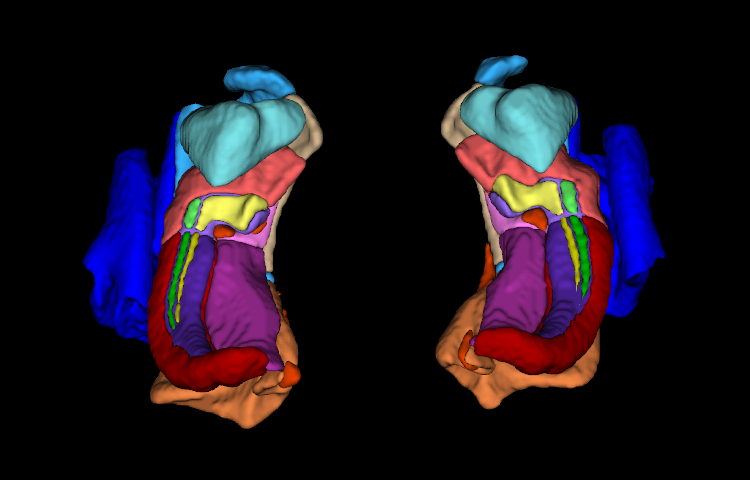
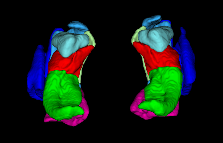
How it works
ASHS atlases are built from manual segmentations of T2-weighted MRI with thick slices. HyperASHS first turns each atlas subject into an isotropic training example. An implicit neural representation (INR) is fitted jointly to the subject's T2w MRI, T1w MRI and manual segmentation, and then sampled on a fine isotropic grid. An nnU-Net model is then trained on these upsampled images. When you segment a new subject, the neck is trimmed from the T1w scan, the T1w and T2w scans are registered to each other and to a template, regions of interest around each hippocampus are extracted and upsampled, and the trained model labels them directly at isotropic resolution.
flowchart TB
  subgraph T["Training: once per model"]
    direction LR
    A["Atlas subjects:<br/>T2w + T1w MRI,<br/>manual segmentation"] --> B["INR upsampling<br/>to isotropic<br/>resolution"] --> C["nnU-Net training<br/>with modality<br/>augmentation"]
  end
  subgraph S["Segmentation: hrashs run"]
    direction LR
    D["New subject:<br/>T2w + T1w MRI<br/>or T1w only"] --> E["Neck trimming,<br/>registration,<br/>MTL ROI extraction"] --> F["Isotropic<br/>nnU-Net<br/>inference"] --> G["Segmentations,<br/>volumes,<br/>QC images"]
  end
  T -- "pre-trained model" --> S
In the paper describing the method, thickness and volume measures from the isotropic model separated people with mild cognitive impairment from cognitively unimpaired people with larger effect sizes than models trained on anisotropic data, and were more stable in test-retest scans.
Available models
| Model ID | Input MRI | Structures | Population |
|---|---|---|---|
PMC_Atlas |
T2w (~0.4 × 0.4 × 2.0 mm³) + T1w (~1 mm³) | Hippocampal subfields, anterior MTL, amygdala, ERC, BA35, BA36, PHC | Older adults, neurodegenerative disease |
PMC_T1_Atlas |
T1w only (~1 mm³) | Anterior/posterior hippocampus, amygdala, ERC, BA35, BA36, PHC | Older adults, neurodegenerative disease |
HSG_Body_Atlas |
T2w + T1w | Hippocampal subfields in the hippocampal body (HSG harmonized protocol) | See hrashs desc HSG_Body_Atlas |
Run hrashs list to see the current list of models, and hrashs desc <model_id> for details. See the Quick Start to choose one.
Getting started
-
Pick a model, check your MRI data and run your first segmentation.
-
Send your scans to our server from ITK-SNAP. No installation needed.
-
Install HyperASHS from PyPI on a Linux workstation with an NVIDIA GPU.
-
Run
hrashs runand understand the outputs and QC images. -
Train HyperASHS on your own manual segmentation protocol.
About us
HyperASHS is developed by members of the Penn Image Computing and Science Laboratory (PICSL) in the Department of Radiology at the University of Pennsylvania. The source code is on GitHub.
Using classic ASHS? See the equivalent page for ASHS Classic.